Shoulder Muscles Diagram Back
Shoulder Muscles Diagram Back. 5 exercises to improve scapular stabilization and prevent elbow. This diagram depicts back shoulder muscles. Shoulder flexion is movement of the shoulder in a forward motion. The following diagram shows all the major back muscles. Around the shoulder, muscles in the back, neck, shoulder, chest and upper arm all work together to support and move the shoulder. Some of these muscles are quite large and cover broad areas.
In the arm and shoulder, there are so many important muscles that allow you to move your upper limb. Human anatomy lovely shoulder muscle diagram shoulder muscles. Although three ligaments protect and surround the shoulder joint, most of its stability comes from the powerful muscles and tendons of the rotator cuff. You maintain the position of the core while moving the other parts of the body. Muscles of the back can be divided into superficial, intermediate, and deep group.since the all the back muscles originate in embryo (fetus) form by locations other than the back, muscles in the.

Understanding shoulder pain neuromuscular therapy of vermont.
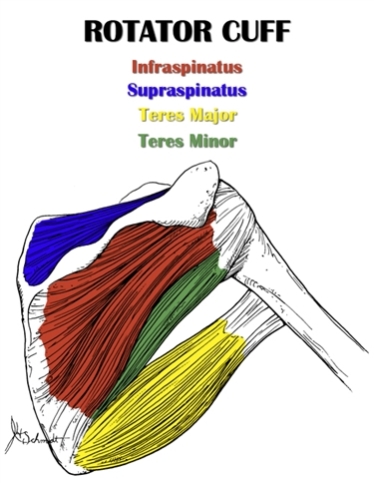
The anterior deltoid, the lateral deltoid, and the posterior deltoid. Published march 30, 2018 at 1000 × 871 in shoulder muscles diagrams. As one of the four muscles of the rotator cuff, the main function is to externally rotate the humerus and stabilize the shoulder joint. Understanding shoulder pain neuromuscular therapy of vermont. Within this group of back muscles you will find the latissimus dorsi, the trapezius, levator scapulae and the rhomboids. Whilst the superficial muscles of the back allow movements at the shoulder, the intermediate muscles of. Weak muscle tissues are amongst the prime causes of back discomfort, especially in the decrease back. The human back extends from the buttocks to the posterior portion of the neck and shoulders. The deltoid, teres major, teres minor, infraspinatus, supraspinatus (not shown) and subscapularis muscles (not shown) all extend from the scapula to the humerus and act on the shoulder joint. It is one of the main muscles that. Ready to test your knowledge on those muscles?
Around the shoulder, muscles in the back, neck, shoulder, chest and upper arm all work together to support and move the shoulder. Within this group of back muscles you will find the latissimus dorsi, the trapezius, levator scapulae and the rhomboids. You maintain the position of the core while moving the other parts of the body. What's important to note here is that from the back it swings under the triceps and. Shoulder muscles, shoulder muscles name, shoulder muscles pain, shoulder muscles workout. Ready to test your knowledge on those muscles?
One of the most important of these for shoulder motion is the deltoid.
Published march 30, 2018 at 1000 × 871 in shoulder muscles diagrams. An example of shoulder flexion can be seen when reaching forward to grasp an object. The latissimus dorsi muscle spans from the lower back to the upper arm and is partially covered by the trapezius. Anatomy of the upper back youtube. It is one of the main muscles that. Each muscle of the shoulder assists with specific movements. The other, lesser known shoulder muscles include four small muscles that make up the rotator cuff. The deltoid, teres major, teres minor, infraspinatus, supraspinatus (not shown) and subscapularis muscles (not shown) all extend from the scapula to the humerus and act on the shoulder joint. Understanding shoulder pain neuromuscular therapy of vermont. Ankle muscles diagram, back muscles diagram, chest muscles diagram, diagram of shoulder muscles and tendons, hip muscles diagram, knee muscles diagram, neck muscles diagram, rotator cuff muscles diagram, human muscles, ankle muscles diagram, back muscles diagram. Shoulder muscles, shoulder muscles name, shoulder muscles pain, shoulder muscles workout.
Other muscles are small and cover much less space. The shoulders are called the deltoid muscles or the deltoids. Human anatomy diagrams show internal organs, cells, systems, conditions, symptoms and sickness information and/or tips for healthy living. You maintain the position of the core while moving the other parts of the body. Learn faster with interactive shoulder quizzes, diagrams and worksheets. Diagram of upper back muscles and human shoulder muscle diagram.

The shoulder muscles produce the characteristic shape of the shoulder and can be classified into two groups:
Ready to test your knowledge on those muscles? Around the shoulder, muscles in the back, neck, shoulder, chest and upper arm all work together to support and move the shoulder. Other muscles are small and cover much less space. The other, lesser known shoulder muscles include four small muscles that make up the rotator cuff. Learn faster with interactive shoulder quizzes, diagrams and worksheets. There are three sections to the shoulder muscle: The muscles of the shoulder bridge the transitions from the torso into the head/neck area and into the upper extremities of the arms and hands. Start studying back & shoulder muscles. The shoulder muscles produce the characteristic shape of the shoulder and can be classified into two groups: Human anatomy diagrams show internal organs, cells, systems, conditions, symptoms and sickness information and/or tips for healthy living. The anterior deltoid, the lateral deltoid, and the posterior deltoid. Together these are known as the rotator cuff muscles. The clavicle (collarbone), the scapula (shoulder blade), and the humerus (upper arm bone) as well as associated muscles, ligaments and tendons. The next life study seated female figure, shows the upper part of the pectoralis major positioned flat against the rib cage, with very little thickness. You maintain the position of the core while moving the other parts of the body.
The back's muscles start at the top of the back (named the cervical vertebrae) and go to the tailbone (also named the coccyx) shoulder muscles diagram. This diagram depicts back shoulder muscles.

Posting Komentar untuk "Shoulder Muscles Diagram Back"